Cell Concentration Calculator Imagej
2A throughout the cell concentration range from very high to very low despite the issue with cells coalescing at higher concentrationsImportantly it took from 35 to 14 min to count the cells manually depending on the dilution factor ie cell concentration. These instructions cover two very basic ways to get cell counts one manual and one.

Object Based Analyses In Fiji Imagej To Measure Local Rna Translation Sites In Neurites In Response To Ab1 42 Oligomers Biorxiv
ImageJ is a freeware image analysis program developed at the National Institutes of Health https.

Cell concentration calculator imagej. Measuring cell fluorescence using ImageJ. This application note describes a method to measure cell migration using ImageJ by counting the number of cells that have migrated into the Detection Zone in an Oris Cell Migration Assay. This action fills in the image width and height text boxes with the image resolution in pixels.
HelloIm very new to ImageJ and any kind of image processing really. Extract the zip file within your ImageJ directory. It is implemented as a plugin for ImageJ image processing and analysis program.
Click on segmentation s. If you wish to perform dilution factor or fold dilution calculations for solutions with cells per volume concentration units eg cellsmL cellsL 10 3 cellsmL 10 6 cellsL etc use our Dilution Factor Calculator - Cells per Volume. Determining the concentration of protein in SDS-PAGE gel bands using ImageJ To determine protein concentration you will need to have a standard curve to compare your samples to - For 5GB1 BSA works great as a protein standard and a range of 0025 gL to 50 gL works well as a range for the standard curve.
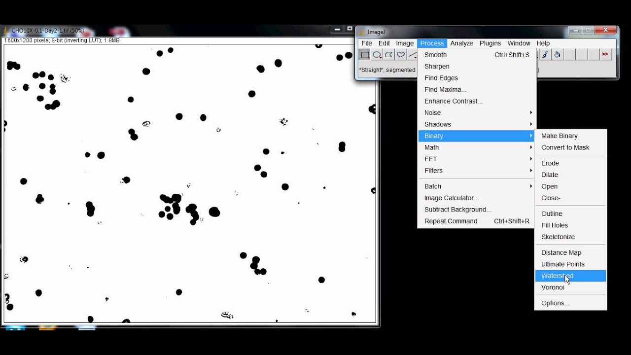
Change thresholding method to Default. Cell Concentration Calculator Transwell Counter. Adipocyte Cell Counting.
ImageJ plugin for Cell Segmentation. Use this formula to calculate the corrected total cell fluorescence CTCF. This software is designed to process transmission electron microscopy images containing cells.
Select freehanding tool to outline any cells that were not counted. The two algorithms showed remarkable consistency with each other and with the manual cell count Fig. In this interactive tutorial students will get acquainted with the applications of ImageJ.
Use 40-40000 for size typically. Image1 or both Image1 and Image2 can be stacks. The plugins files will be placed within the correct directories.
In ImageJ under the cell concentration calculator open the image and click on the Get Image Dimension button. Open image of interest. With macro installed click on segmentation.
Performs arithmetic and logical operations between two images selected from popup menus. Modern microscopy techniques generate an enormous amount of data in the form of images. Next select the straight line tool and draw a straight line across the entire length of the hemocytometers primary p-square by clicking.
Two Ways to Count Cells with ImageJ Figuring out how many cells are in an image is a common need in image analysis. Support is available on the mailing list and on the imagesc forum. What does the Image Calculator command do.
To extract the numerical data from the images a free and user-friendly software called ImageJ is available at the NIH website. May not work correctly after using Load Markers to load more than 8 counter types from an XML file. Counting Cells in Migration Assays with ImageJ.
Segmenting Cells for Area Calculation. CTCF Integrated Density. There are several ways to go about this some more involved than others.
Meant to be used in both the teaching and research laboratory this calculator see below can be utilized to perform dilution factor calculations when working with solutions having cells per volume ie cells over volume concentration units such as cellsmL cellsL 10 3 cellsmL 10 6 cellsL etc. It contains semiautomatic tools for segmentation of. Notice that rounded up mitotic cells appear to have a much higher level of staining due to its smaller size concentrating the staining in a smaller space.
If both are stacks they must have the same number of slices. Manual analysis of these images produces biased results that are often not reproducible. Posted by 3 days ago.
OBrien J Hayder H Peng C. 2B but it took only. These calculations are commonly performed when working with culture media containing.
Segmenting Cells for Area Calculation. I am having some trouble getting ImageJ to reliably recognize the cells. Image1 and Image2 must be the same data type but they do not have to be the same size.
These calculations are commonly performed when working with culture media containing living cells such as bacterial cells or mammalian cells. Automated Quantification and Analysis of Cell Counting Procedures Using ImageJ Plugins. Download cell_counterjar to the plugins folder or subfolder restart ImageJ and there will be a new Cell Counter command in the Plugins menu or submenu.
Set boundaries for sizing of cell.
Counting Cells With Imagej Youtube

Cell Density Map Image Analysis Image Sc Forum
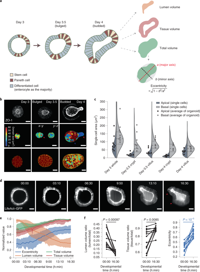
Cell Fate Coordinates Mechano Osmotic Forces In Intestinal Crypt Formation Nature Cell Biology
Regulation Of Gene Transfection By Cell Size Shape And Elongation On Micropatterned Surfaces Journal Of Materials Chemistry B Rsc Publishing

Object Based Analyses In Fiji Imagej To Measure Local Rna Translation Sites In Neurites In Response To Ab1 42 Oligomers Biorxiv
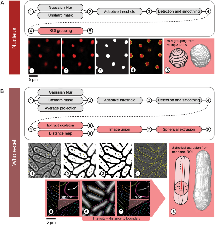
Pomegranate 2d Segmentation And 3d Reconstruction For Fission Yeast And Other Radially Symmetric Cells Scientific Reports

Overriding Native Cell Coordination Enhances External Programming Of Collective Cell Migration Pnas

Pin On This Is My Research Board

Pdf Calculate The Corrected Total Cell Fluorescence Ctcf

Fiji For Quantification Cell Segmentation Youtube
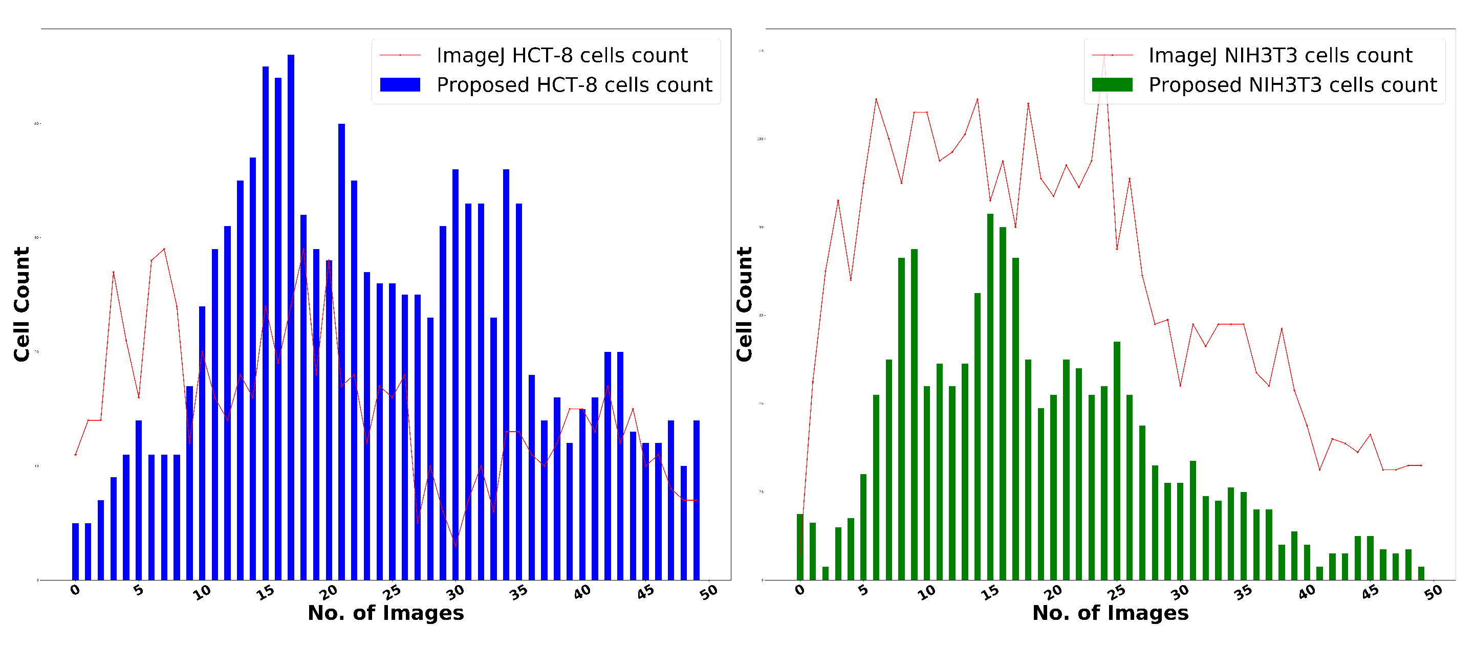
Applied Sciences Free Full Text Quantitative And Qualitative Image Analysis Of In Vitro Co Culture 3d Tumor Spheroid Model By Employing Image Processing Techniques Html
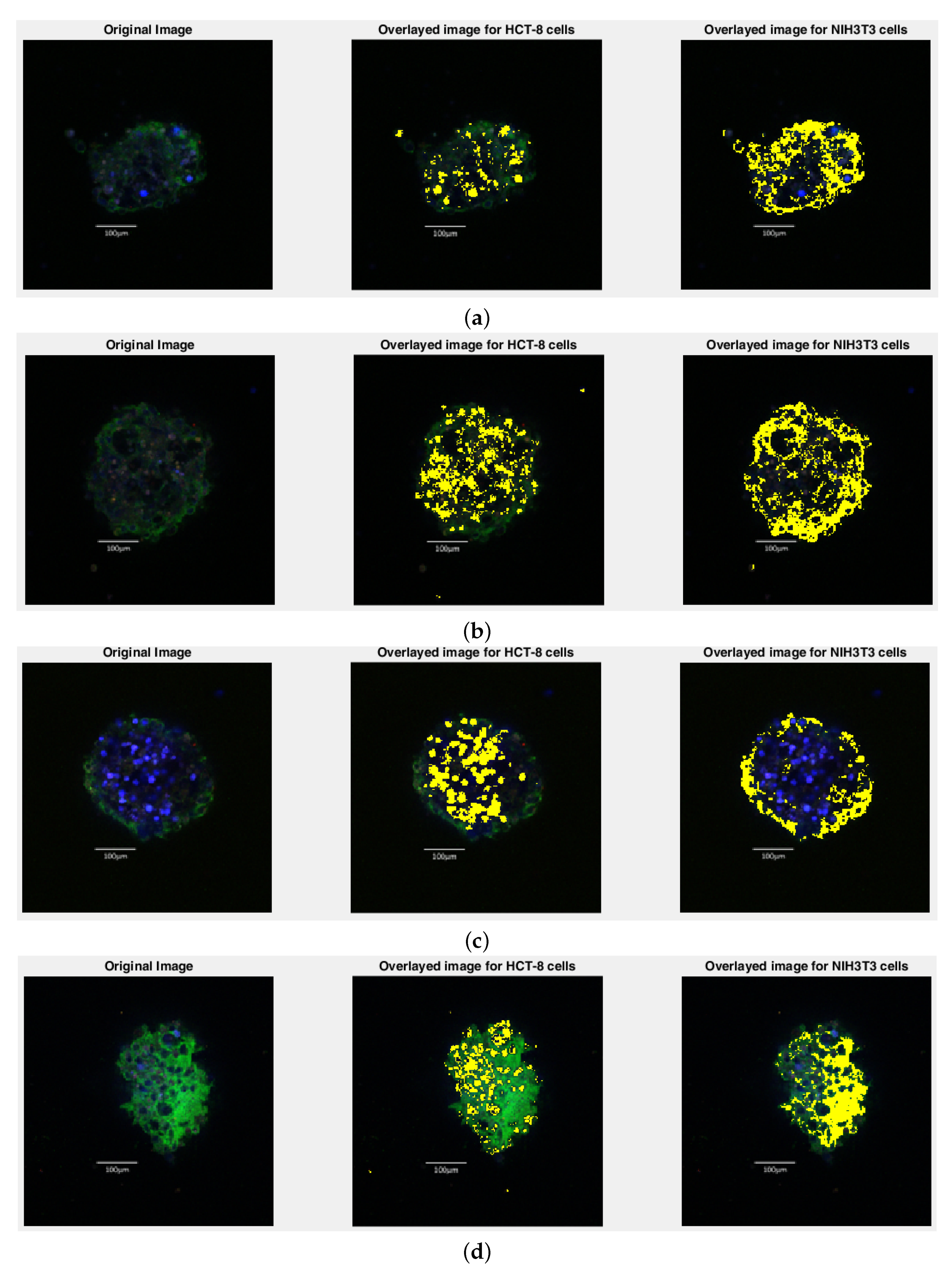
Applied Sciences Free Full Text Quantitative And Qualitative Image Analysis Of In Vitro Co Culture 3d Tumor Spheroid Model By Employing Image Processing Techniques Html

Cell Culture Media A Review Cell Culture Medium Culture Media Media

Regulation Of Gene Transfection By Cell Size Shape And Elongation On Micropatterned Surfaces Journal Of Materials Chemistry B Rsc Publishing

Automated Quantification And Analysis Of Cell Counting Procedures Using Imagej Plugins Abstract Europe Pmc
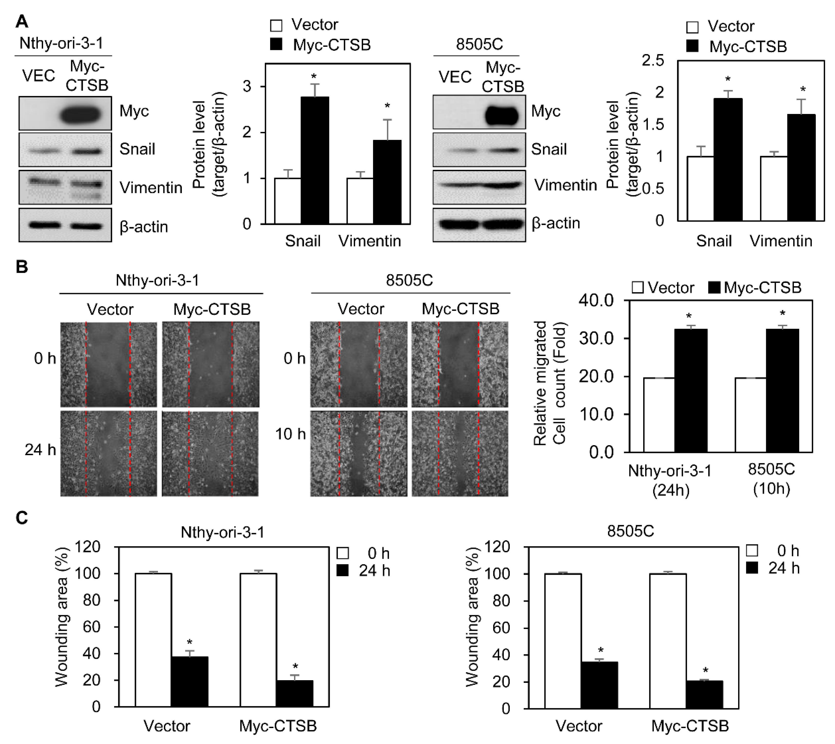
Ijms Free Full Text Clinicopathologic Analysis Of Cathepsin B As A Prognostic Marker Of Thyroid Cancer Html

Spattrack An Imaging Toolbox For Analysis Of Vesicle Motility And Distribution In Living Cells Lund 2014 Traffic Wiley Online Library
Plant Egg Cell Fate Determination Depends On Its Exact Position In Female Gametophyte Pnas
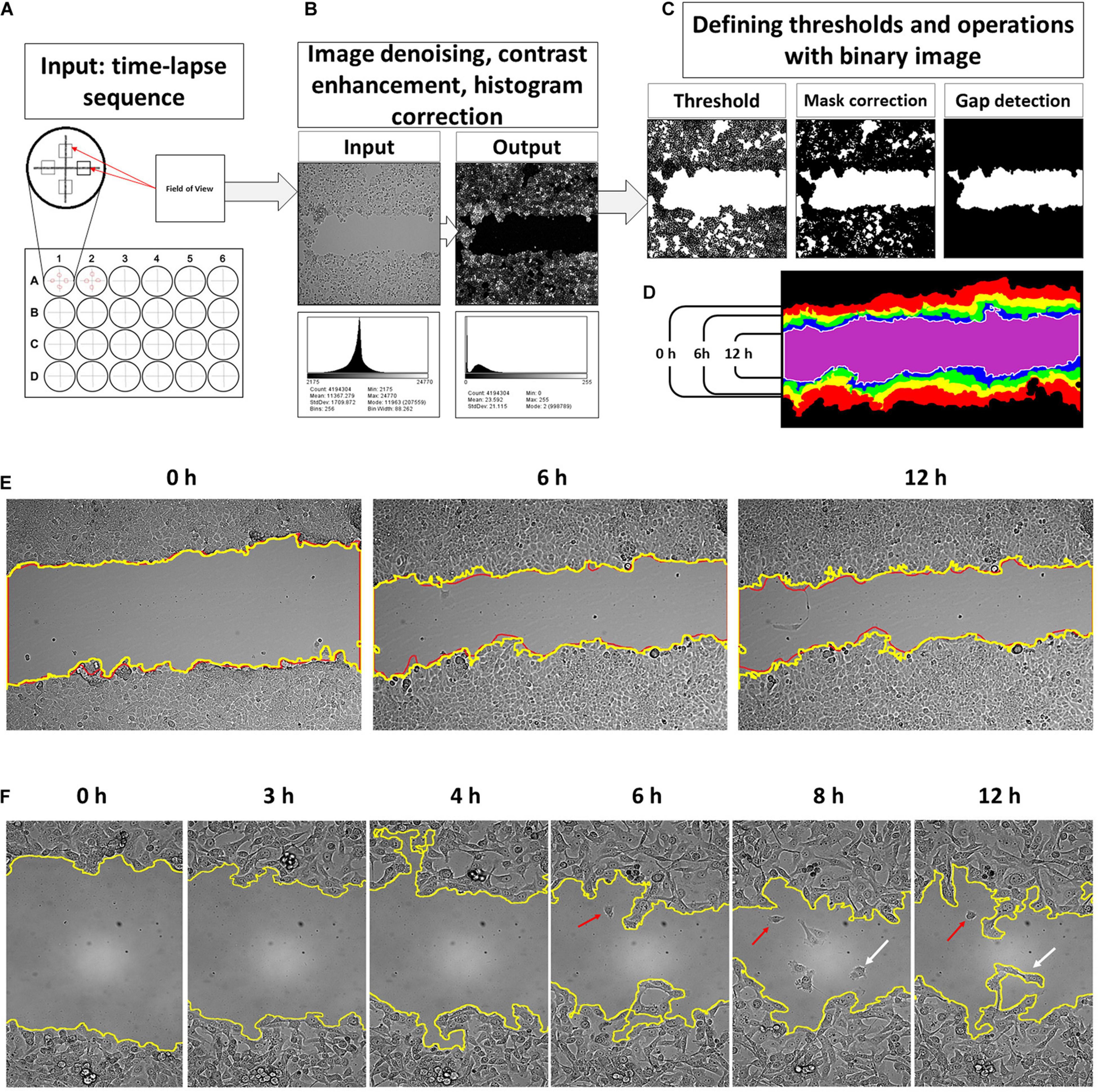
Frontiers The Frequent Sampling Of Wound Scratch Assay Reveals The Opportunity Window For Quantitative Evaluation Of Cell Motility Impeding Drugs Cell And Developmental Biology
Post a Comment for "Cell Concentration Calculator Imagej"